Category Archives: Light Microscope

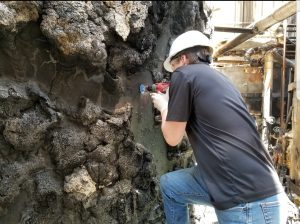
Sample preparation of steel tanks.


Sometimes projects involve samples or artifacts too large or too valuable to remove or cut for evaluation in MEE’s laboratories. In these cases, in situ evaluation is required.

For this project, MEE was asked to evaluate the functional integrity of large steel tanks that had been exposed to elevated temperatures from a fire. Associate Engineer, Henry Ahrenholtz, is seen here preparing a field metallographic specimen. The tank surface was prepared by grinding, polishing and chemical etching to reveal the microstructure of the tank material. A replica of the prepared surface was made and then examined using a portable microscope. A Materials Engineer’s specialized knowledge of the material properties and behavior under certain environmental conditions combined with metallographic expertise in the field provided valuable information for the client to aid in assessing any damage to the tanks caused by fire.



Microscope Monday

posted January 2020
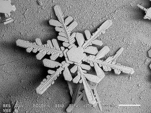
SEM image of snowflake

Snowflake

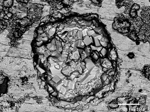
Corrosion Pit

Copper corrosion pit

Twenty years ago, MEE Lab Manager, Kurt Schenk, figured out a way to capture the beauty of a freshly fallen snowflake with an SEM. Those images and hundreds of others taken over the years in our laboratory have been saved in a file on our network simply called “cool images”.

Five years ago, we thought it would be fun to share some of our favorite cool images on social media and the MEE Facebook page was created. We have since posted over 180 images. Some are just-for-fun, like snowflakes, insects and even the grooves in an LP, but most of the images are the kind of things we observe in our daily work. We have posted images that highlight fracture mechanisms, unique microstructures and particular imaging techniques used to gather critical data needed in a failure analysis investigation.

We weren’t familiar with the Minneapolis city flag until just recently, but were pleasantly surprised to see one of the symbols on it is a microscope. That seems fitting to us! Microscopes are one of the most important tools we use at MEE to provide our clients in the medical device, industrial and electronics industries with high-quality materials characterization for their advanced materials and critical components.

MEE has three scanning electron microscopes (and a new one coming this summer) and a light microscopy laboratory equipped with a variety of light microscopes with magnifications ranging from 5X to 2,400X.

There is a small contingent of Minneapolis citizens advocating for an update of the flag. We would hate to see the microscope image go away but either way, MEE will always be flying the microscopy banner.


City of Minneapolis Flag

Light Microscopy

posted December 2016

Our laboratory at  MEE has a variety of light microscopes with magnifications ranging from 5X to 2400X. Light microscopes aid in low-magnification observation of sample surface features using visible light. The Keyence VHX 1000 is a state-of-the-art digital microscope that captures images with an extended depth of field and allows for 3D measurements of surface features. The laboratory also has a range of stereo microscopes for low-magnification inspection and imaging. The MEE laboratory staff know how to properly prepare and examine samples to obtain the optimum materials characterization data.

Contact us to discuss which evaluation method is best suited to solve your materials-related problems.

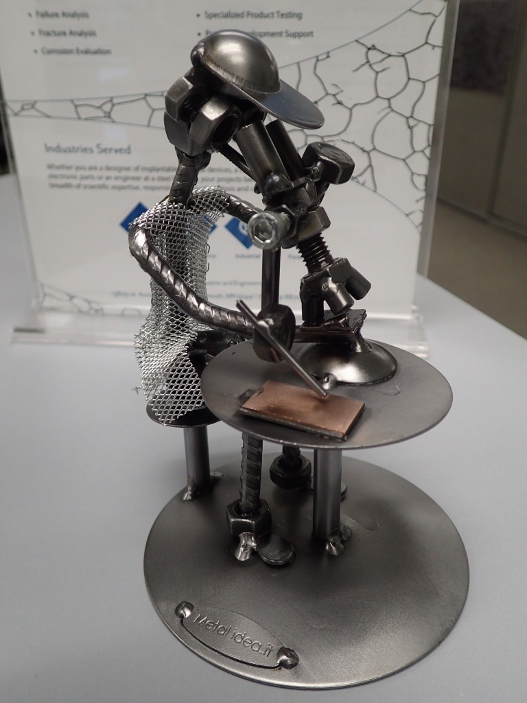
Metal sculpture

One of our staff found this metal sculpture on his recent vacation to Italy. Our newest metallurgical microscopist is always on the job. 

Filed under: About MEE, Light Microscope,

Another Way to Look at Things…

posted December 2015

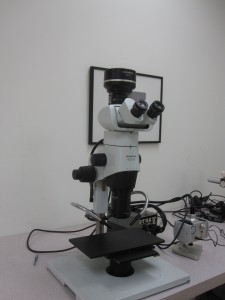
We just purchased a research grade Olympus SZX16 stereo microscope equipped with a super high resolution, digital, micro-imaging camera.  This is an excellent addition to our light microscopy laboratory. With easy shifting from macro-view to micro-view,  it is a specialized and powerful tool for fracture examination, corrosion studies and general failure analysis.

We’re looking forward to putting our newest microscope to work.

Olympus SZX16 stereo microscope with digital camera.

Olympus SZX16 Stereo Microscope with digital camera.

MEE on Facebook

posted June 2015

FB_FindUsOnFacebook-114


MEE, Inc. has been in business since 1995 and has captured tens of thousands of images using scanning electron microscopes and light microscopes. Our technical staff has long kept a file of “cool” images which we are sharing on our new Facebook page.  

Like our page and enjoy some interesting and unique images. Facebook posts will be updated on average two to three times a month.